PPT Abdominal wall PowerPoint Presentation ID295293
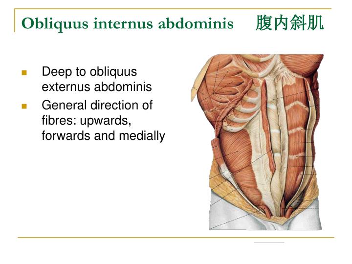
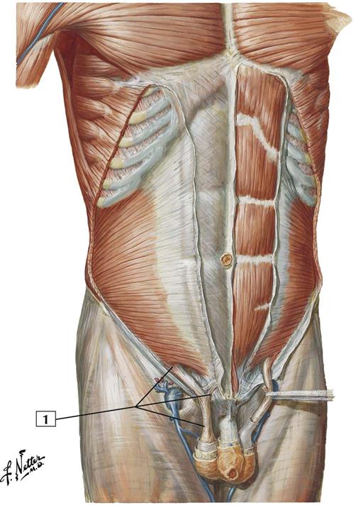
Musculus obliquus internus abdominis 1/5. Synonyms: none. The aponeuroses of internal oblique, external oblique and transversus abdominis muscles extend vertically across the anterior aspect of the abdomen. While crossing over to the opposite side, their aponeurotic fibers interweave along the midline of the abdomen, forming the thickening.
M. obliquus externus abdominis Anatomie und Funktion Kenhub
A video and a drawing of the external abdominal oblique muscle. Anatomical structures in item: Abdomen. Musculus obliquus externus abdominis. Musculus obliquus internus abdominis. Musculus transversus abdominis. Musculus rectus abdominis. Fascia abdominis. Fascia investiens intermedia abdominis.
1942 Obliquus Internus Abdominis Transversus Abdominis Original Vintage Print Anatomy
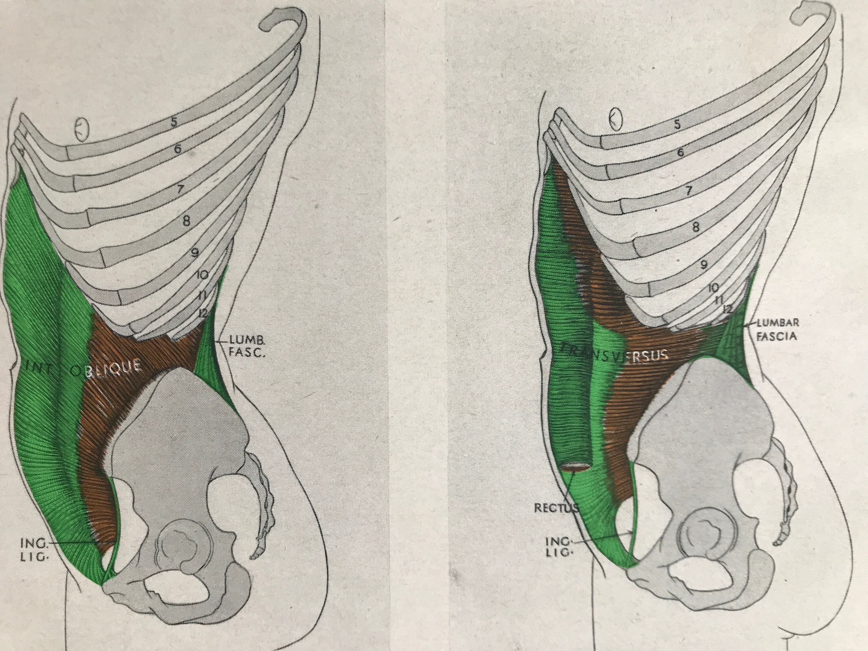
Description: The Obliquus internus abdominis (Internal or ascending oblique muscle), thinner and smaller than the Obliquus externus, beneath which it lies, is of an irregularly quadrilateral form, and situated at the lateral and anterior parts of the abdomen.
Musculus obliquus internus abdominis Anatomie Kenhub
m the following regression model: Yt m quasi-Poisson (nt t) log (t) = b0 + log (nt) + s (t, 8 df/year) + b1 dayst + b2 holi-dayst + Age, b3 summerpopt + cb (tempt) wherein < temp is now the maximum temperature modeled by a distributed lagnonlinearmodel.29 Thistypeofmodelissuitablefor describing the complex nonlinear time-lag dependencies.

M. obliquus internus YouTube
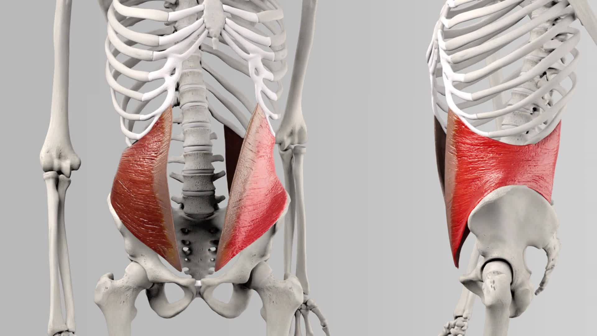
Der Musculus obliquus internus abdominis (innerer schräger Bauchmuskel) wird zu den seitlichen Bauchmuskeln gezählt. Er befindet sich flächig zwischen dem M. transversus abdominis (querer Bauchmuskel) und dem M. obliquus externus abdominis (äußerer schräger Bauchmuskel), dessen Fasern im Gegensatz zu seinen eigenen in einem 90° Winkel verlaufen.
Základy sportovní kineziologie Fakulta sportovních studií
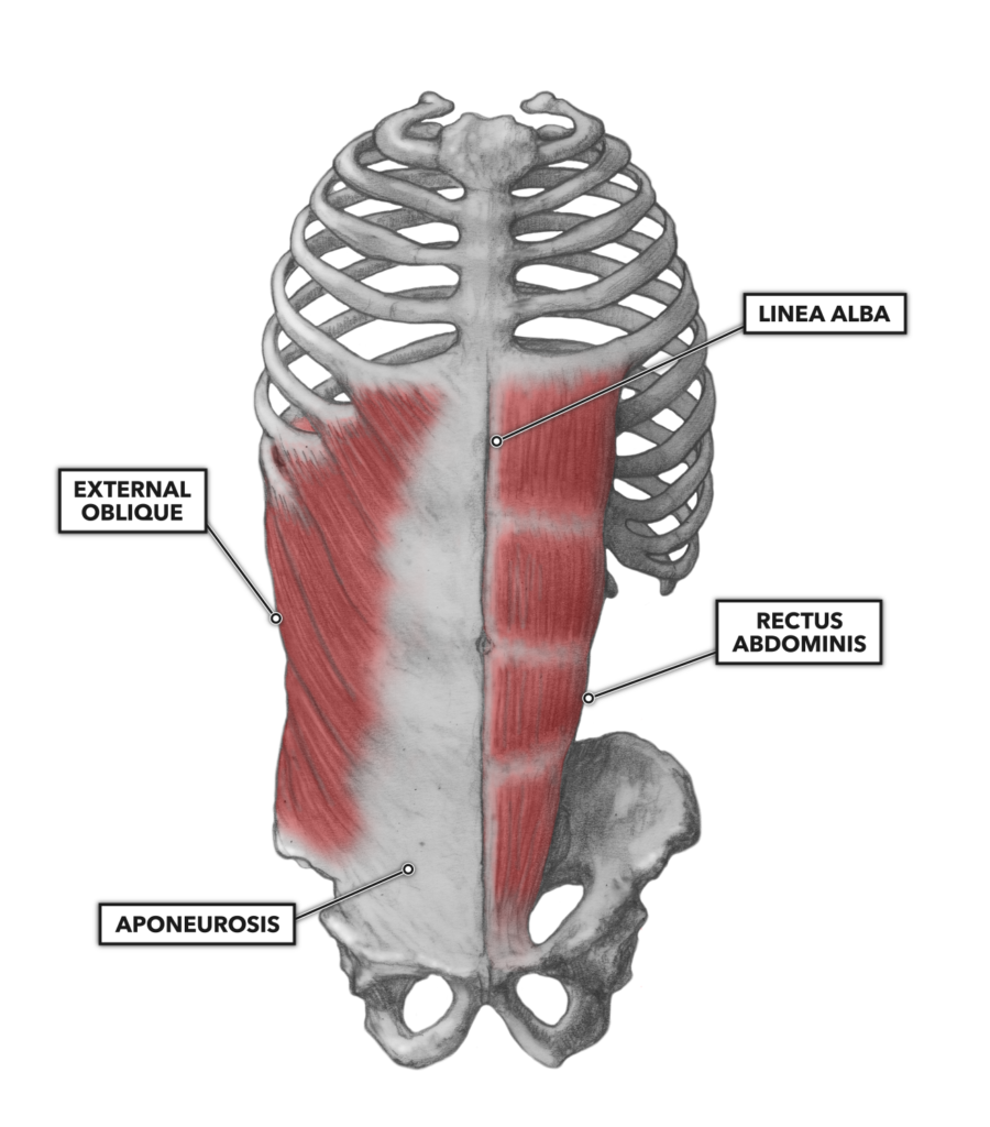
The Obliquus externus abdominis ( External or descending oblique muscle ), situated on the lateral and anterior parts of the abdomen, is the largest and the most superficial of the three flat muscles in this region. It is broad, thin, and irregularly quadrilateral, its muscular portion occupying the side, its aponeurosis the anterior wall of.

Musculus obliquus internus abdominis sportbachelor
M. obliquus externus abdominis. M. obliquus internus abdominis. Preferred test of the anterior fibres of the external oblique muscles on the right. Alternative test for the oblique abdominal muscles. Preferred test of the anterior fibres of the internal oblique muscles on the right. Test of the posterior fibres of the oblique abdominal muscles.
Musculus obliquus externus abdominis sportbachelor
transversus abdominis and the m. obliquus internus abdominis for the lateral injections. Minimal dye solution was found in a fascial plane superficial to the TAP, between the mm. obliquus internus abdominis and obliquus externus abdominis or between their aponeuroses, in all 16 lateral injections (Figure 9). The surface of these muscles was.

Anatomy Stock Images torsoabdominalmusclesmusculusobliquesinternusinternaloblique
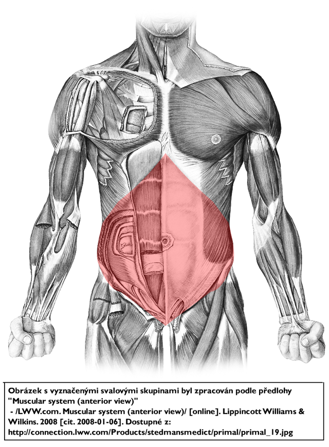
The abdominal internal oblique muscle, also internal oblique muscle or interior oblique, is an abdominal muscle in the abdominal wall that lies below the external oblique muscle and just above the transverse abdominal muscle . Structure
Musculus obliquus internus abdominis DocCheck
Until now, m. obliquus internus abdominis avulsion at the iliac crest has been described only in three Australian football players (two papers). To date, operative treatment of this lesion is not published. We present the first report on a respective operative technique and also describe the rehabilitation. An orthopedic surgeon should be aware.
What Is The Action Of Internal Oblique
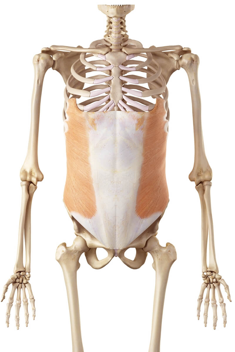
The internal oblique muscle forms part of the superficial covering of the anterolateral abdominal wall. It is one of three large flat muscles in this region that lie under cover of the external oblique muscle.
CrossFit Lumbar Muscles, Part 2
m. obliquus internus abdominis (internal oblique muscle) Muscle Overview: Location, Shape, Function, Insertion, and Origin Location The oblique muscles are located on both sides of the abdomen (sides of waist). External obliques. Top layer, closest to the skin, running downwards and forwards. Internal obliques.

Základy sportovní kineziologie Fakulta sportovních studií
Musculus obliquus internus abdominis Začátek: thorakolumbální fascie; crista iliaca; ligamentum inguinale. Úpon: linea alba; tři poslední žebra; dolní okraj srůstá s aponeurosou musculus transversus abdominis a tvoří tak falx inguinalis. Inervace: nervi intercostales (8.-11.); nervus subcostalis; nervus iliohypogastricus; nervus ilioinguinalis.
Musculus obliquus internus abdominis Anatomie Kenhub
To prevent confusions, be aware that in the textbooks it is uncommon to distinguish layers of the external oblique muscle, hence the internal abdominal oblique is most commonly described as the second, or middle layer of the lateral abdominal wall muscles. The complete video can be seen here.
Internal oblique, external oblique, transversus muscle Kenhub
Internal abdominal oblique is a broad thin muscular sheet found on the lateral side of the abdomen. Going from superficial to deep, the external abdominal oblique, internal abdominal oblique and transversus abdominis comprise the three distinct layers of the lateral abdominal wall.

M. Obliquus Internus Abdominis Innerer Bauchmuskel Ansatz Ursprung Funktion BLizenz Prüfung
Musculus obliquus internus abdominis Quick Facts Origin Insertion Key Features & Anatomical Relations Actions List of Clinical Correlates References Actions Quick Facts Origin: Thoracolumbar fascia, iliac crest, inguinal ligament. Insertion: Inferior margins of tenth to twelfth ribs and adjacent costal cartilages, linea alba, pecten pubis.
